Functional Optical Imaging Laboratory
FOIL is dedicated to developing novel optical imaging technologies for studying the brain
Projects
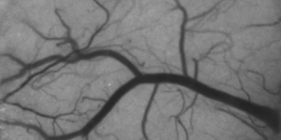
Laser Speckle Contrast Imaging
We develop blood flow imaging systems based on laser speckle contrast imaging (LSCI) to study the effects of ischemic stroke and to better understand the physics of dynamic light scattering.
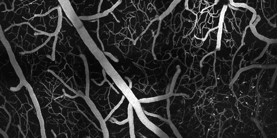
Multiphoton Microscopy
We design and build high-resolution microscopes to study the three-dimensional vascular and neuronal structures of the cortex at depths and resolutions pushing the limits of optical imaging.
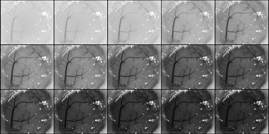
Multi-Exposure Speckle Imaging
We developed an advanced form of LSCI called multi-exposure speckle imaging (MESI) that offers more versatile and robust estimates of blood flow across a wider range of speeds.
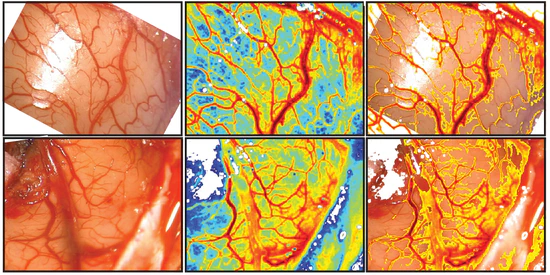
Clinical Speckle Imaging
We are designing and building translational laser speckle contrast imaging systems for clinical applications such as continuous blood flow monitoring during vascular neurosurgery.
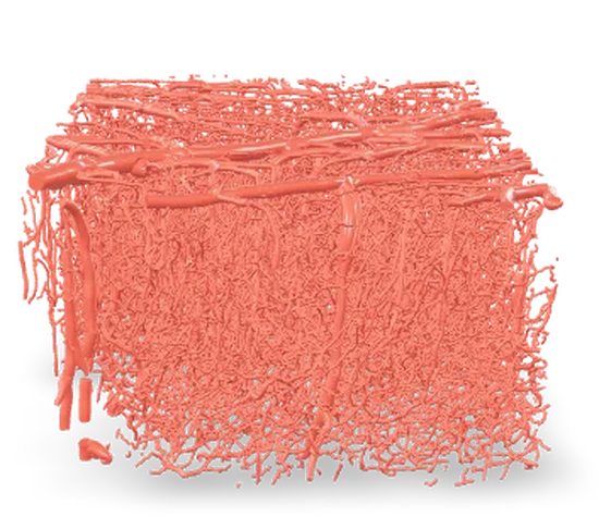
Mouse Blood Vessel AR Models
Explore the intricate structure and function of mouse blood vessels through the lab’s use of Vectorization, Multiphoton Microscopy, and Computational Modeling techniques to create augmented reality models. View our selection of models on your mobile device for an interactive experience.
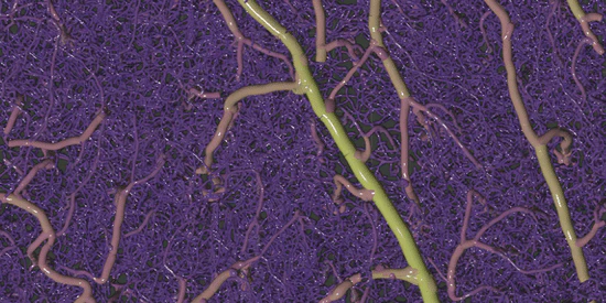
Vascular Vectorization
We developed an automated vectorization method to extract blood vessel geometry and vascular statistics directly from large volumes of unsegmented multiphoton microscopy imagery.
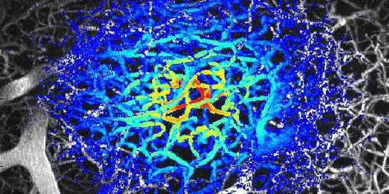
Modeling Light Propagation
We simulate photon propagation through large cortical tissue geometries in order to study the effects of scattering and develop new imaging techniques based on dynamic light scattering.
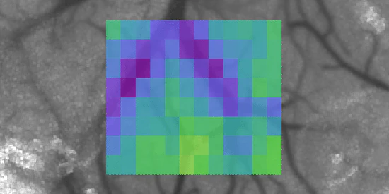
Oxygen Imaging
We developed imaging systems for use with oxygen-sensitive phosphorescent dyes in order to map cortical oxygen tension (pO2) and study its impact on the progression of ischemic stroke.
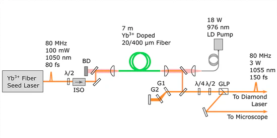
Custom Ultrafast Lasers
We design and build custom ultrafast lasers to push the limits of traditional multiphoton microscopy and enable advanced multi-wavelength excitation strategies.
3D Single Particle Tracking
We built a three-dimensional single particle tracking system capable of spatial localization far beyond the diffraction limit for use in studying molecular transport dynamics.
Contact Information
- adunn@utexas.edu
- 512-232-2808
- Biomedical Engineering Building<br/>107 W. Dean Keeton St., Austin, TX 78723