3D Single Particle Tracking
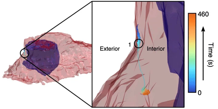 Internalization of single EGFR molecule (Perillo, 2015)
Molecular trafficking within cells, tissues, and engineered three-dimensional (3D) multicellular models is critical to the understanding of the development and treatment of various diseases including cancer. However, current tracking methods are either confined to two dimensions or limited to depths of only 15 µm. Our aim is to develop new 3D tracking techniques and algorithms to improve tracking in tissue and in vivo models. We have recently developed a new tracking method called TSUNAMI capable of quantifying rapid molecular transport dynamics in highly-scattering environments at depths up to 200 µm. The system has a response time of 1 ms with a temporal resolution down to 50 µs in high signal-to-noise conditions, and a spatial localization precision as good as 35 nm.
).](/project/3d-single-particle-tracking/TSUNAMI_hu_b2f96f0c936692c4.webp)
Built on spatiotemporally multiplexed two-photon excitation, this approach requires only one detector for 3D particle tracking and allows for two-photon, multicolor imaging. An initial demonstration of the technique was performed on epidermal growth factor receptor (EGFR) complexes at a depth of 100 µm in tumor spheroids. We are currently working to extend this technique’s capabilities with multi-color detection. Current application areas include early cancer diagnosis in circulating tumor cells, and study of EGFR trafficking in the cytoplasm.