Laser Speckle Contrast Imaging
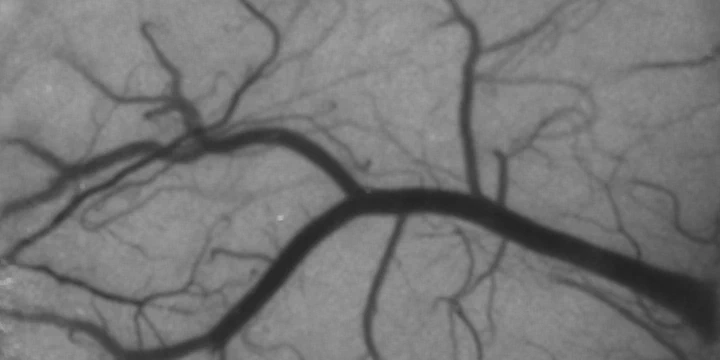 Speckle contrast image of cortical blood flow (Sullender, 2022)
There has been increasing interest in using laser speckle contrast imaging (LSCI) as a tool for imaging blood flow in preclinical research and clinical applications. LSCI utilizes intrinsic tissue contrast from dynamic light scattering to offer a relatively simple technique for visualizing detailed spatiotemporal dynamics of blood flow changes in real-time.
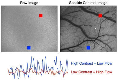
Laser speckle is the random interference pattern produced when coherent light scatters from a medium that can be imaged onto a detector such as a camera. Motion from scattering particles, such as red blood cells in the vasculature, leads to spatial and temporal variations in the speckle pattern. Speckle contrast analysis quantifies the local spatial variance ($K$), or blurring, of the speckle pattern that results from blood flow.
\begin{equation} K = \frac{\sigma_{s}}{\langle{I}\rangle} \end{equation}
Areas with greater motion have more rapid intensity fluctuations and therefore more blurring of the speckle pattern during the camera exposure (Fig. 1). LSCI can be used to quantify relative changes in blood flow, and has been studied both in animal models and in the clinic.
In our lab, we focus on functional brain imaging and use LSCI to study cerebral blood flow (CBF) dynamics. CBF is an important hemodynamic parameter in the brain that can be used to study neurological events such as stroke, cortical spreading depression, and functional activation. We use LSCI in animal models as a tool to better understand the neurophysiological mechanisms behind these events. In the clinic, LSCI is being leveraged as a non-invasive monitoring tool for neurosurgery that could help reduce the risk of postoperative blood flow deficits.
).](/project/laser-speckle-contrast-imaging/webcam_hu_158921102268d2f9.webp)
).](/project/laser-speckle-contrast-imaging/PID.gif)