Multi-Exposure Speckle Imaging
 Speckle contrast images acquired at different exposures
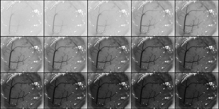
Multi-exposure speckle imaging (MESI) is an advanced form of laser speckle contrast imaging that offers more robust estimates of flow across a wider range of flow speeds. By revisiting the underlying physics behind LSCI to better account for the presence of static scatterers and capturing imagery at multiple exposure times, MESI enables the chronic tracking of cerebral blood flow. These improvements have allowed us to study the hemodynamics of ischemic stroke across timescales spanning seconds to months after the infarct.
).](/project/multi-exposure-speckle-imaging/stroke_hu_5b1c606af80bbff5.webp)