Oxygen Imaging
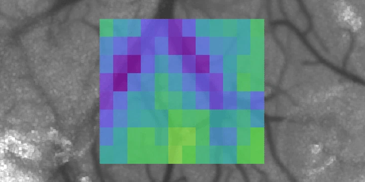 Vascular oxygen tension map (Sullender, 2018)
We use oxygen-sensitive porphyrin probes for noninvasive, highly-sensitive optical oxygenation measurements based on phosphorescence quenching. While an injection of the dye is required, absolute oxygen tension ($p_{\ce{O2}}$) can be directly calculated from the lifetime ($\tau$) of the measured phosphorescence using the Stern-Volmer relationship:
\begin{equation} \frac{I_0}{I} = 1 + k_q\tau_0[Q] \end{equation}
In order to localize the phosphorescent signal, we use either two-photon excitation or structured illumination with a digital micromirror device (DMD). The latter is combined with laser speckle contrast imaging for multimodal imaging of cortical hemodynamics (cerebral blood flow + $p_{\ce{O2}}$). These systems are used to study both the acute and chronic dynamics of ischemic stroke.
).](/project/oxygen-imaging/pO2_roi_hu_fc835628ba4548e2.webp)
).](/project/oxygen-imaging/2P_pO2_hu_2f2e185cfb0468a7.webp)